Entries from December 1, 2013 - December 31, 2013

Thursday
Dec262013

Crust Fungus

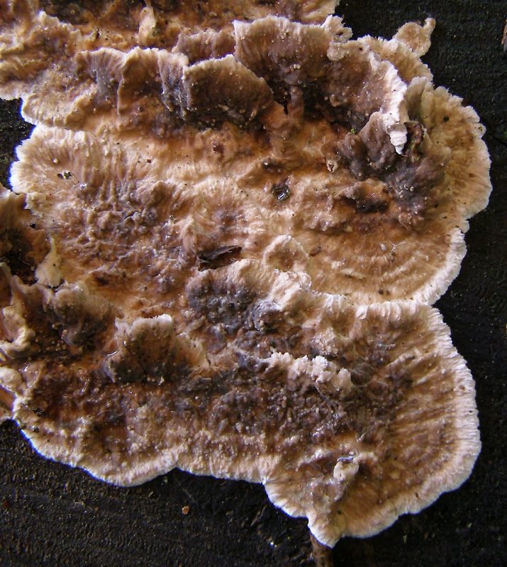
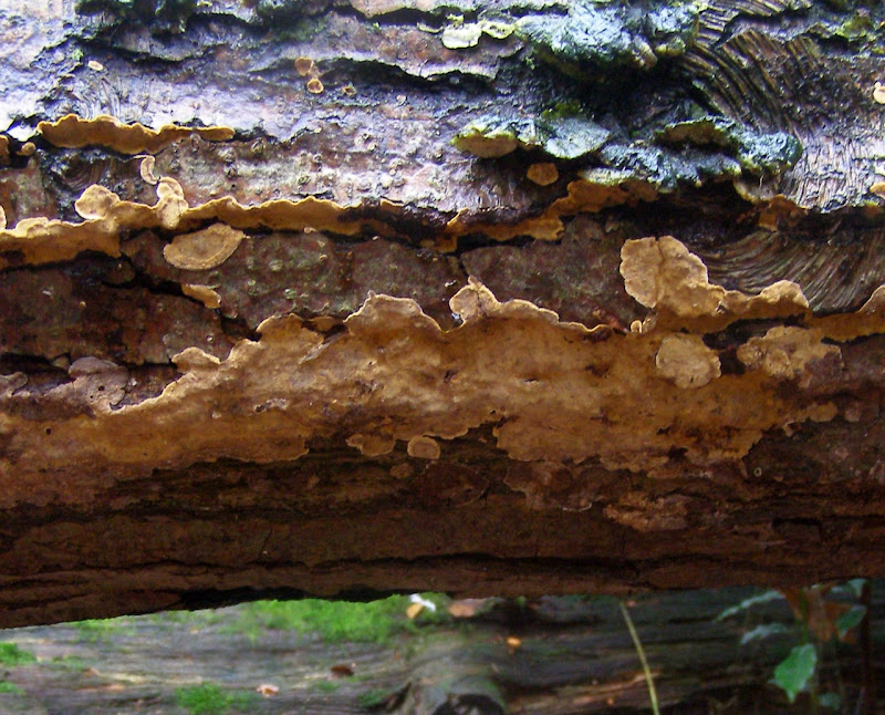
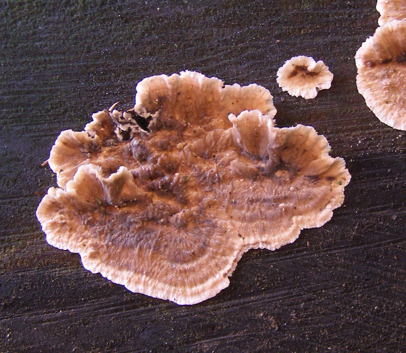
Crust fungus Stereum sanguinolentum growing on the cut end of a conifer log.

Back in March while taking a short cut from my usual walk, I noticed this interesting-looking crust fungus growing on a conifer log near Wessex Hall.  Initially I thought it might be a brownish variant of Chondrostereum purpureum but the sample I took was too dried out to give any spores and didn't show any other distinguishing features so I didn't pursue the matter any further.

Yesterday morning I happened to be passing that way again and noticed the fungus was still there so I took these photos.

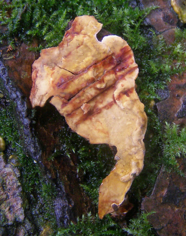
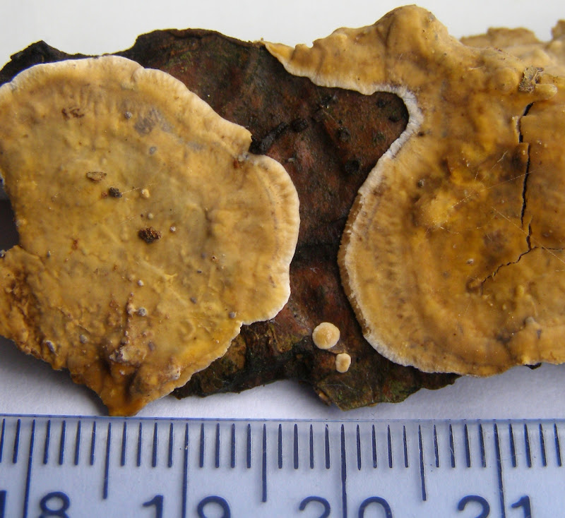
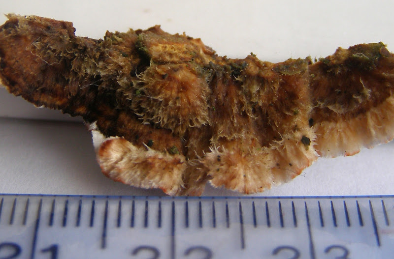
I also took a sample of one of its brackets.  The upper surface of the bracket is hairy:

The lower surface is smooth and lacks any visible pores:

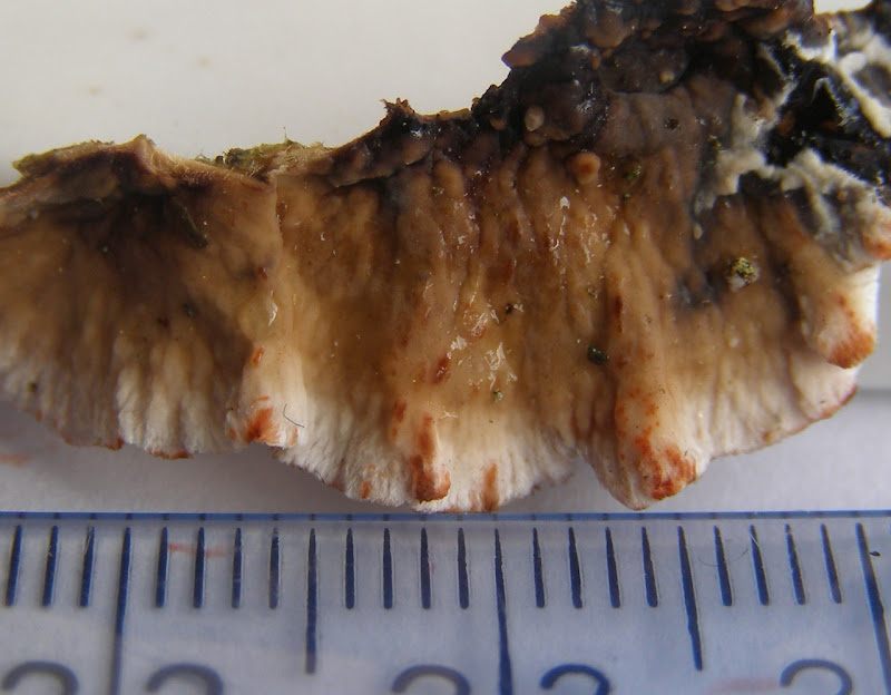
Note also that where the lower surface has been accidentally bruised it has turned red.  The only crust fungus that bleeds red and grows on conifers is Stereum sanguinolentum.  The above sample also bled when I deliberately cut it. The sample I took back in March must have been too dried out to even bleed.

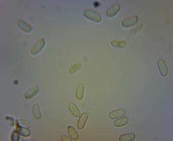
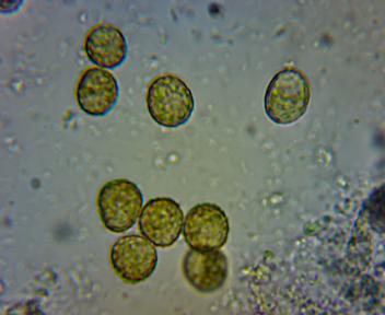
When left 24 hours over a microscope slide the lower surface gave the following spores (magification x600, field width about 86um):

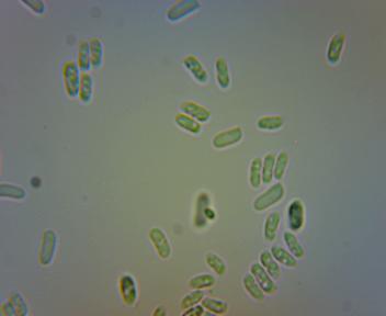
These spores are about 6.5 x 2.5um which is at the lower end of the range (6-)7-10(-10.5) x (2-) 2.5-3(-3.5) um given for Stereum sanguinolentum at this page at MycoBank.

[Note added 2013-12-30: the log on which it was growing was most probably cut from a nearby cedar (Cedrus libani).]

First 2 photos and specimen taken in Whiteknights Park, Reading, UK, on 2013-12-25.

Wednesday
Dec252013

Leaf Mines on Holly

Leaves of holly (Ilex aquilfolium) with leaf mines, probably caused by larvae of the fly Phytomyza ilicis (Diptera: Agromyzidae).  For more info see this page at the Natural History Museum.

Leaves taken in the Wilderness, Whiteknights Park, Reading, UK, on 2013-12-18.

Tuesday
Dec242013

Crust Fungus

On the underside of a fallen birch trunk: a yellowish crust fungus, probably Stereum gausapatum.

It bleeds red liquid hen its surface is cut:

I took the following specimen home to look for spores:

And this it what it gave (x600 magnification, field width about 86um):

These spores are about 8 x 3.5um.  This is a bit smaller than the 9-13 x 3.5 - 5um given for Sterum rugosum at Rogers Mushrooms but nicely matches the 7-8 x 3-3.5um for S. gausapatum given by the same source. 

Apparently, S. sanguinolentum has spores the same size as gausapatum but that species grows only on conifers, not on birch.

First 2 photos and specimen taken in the Wilderness, Whiteknights Park. Reading, UK, on 2013-12-21.

Monday
Dec232013

Slime Mould

Sporangia of a slime mould, probably Trichia decipiens, growing on a decaying log.

I took the following specimen to look at at home:

I squashed one of the sporangia onto a microscope slide but was unable to find any spores so I left the sample to mature. 

After a couple of days the sporangia had turned a dark olive green:


Under the microscope they yielded the following spores (x600):

The field width is about 86um so the spores are about 10 x 11um.

Also present were the long tapered elaters characteristic of Trichia decipiens:

First photo and specimen taken in the Wilderness, Whiteknights Park, Reading, UK, on 2013-12-18.

Sunday
Dec222013

Lacewing

A green lacewing, probably a member of the Chrysoperla carnea group of species (Neuroptera: Chrysopidae).  I noticed on our bedroom window at the end of a warm day in mid December.  I trapped it in order to photograph it in daylight defore releasing it again.

Specimen taken in Reading, UK, on 2013-12-17.