Monday
Sep232013
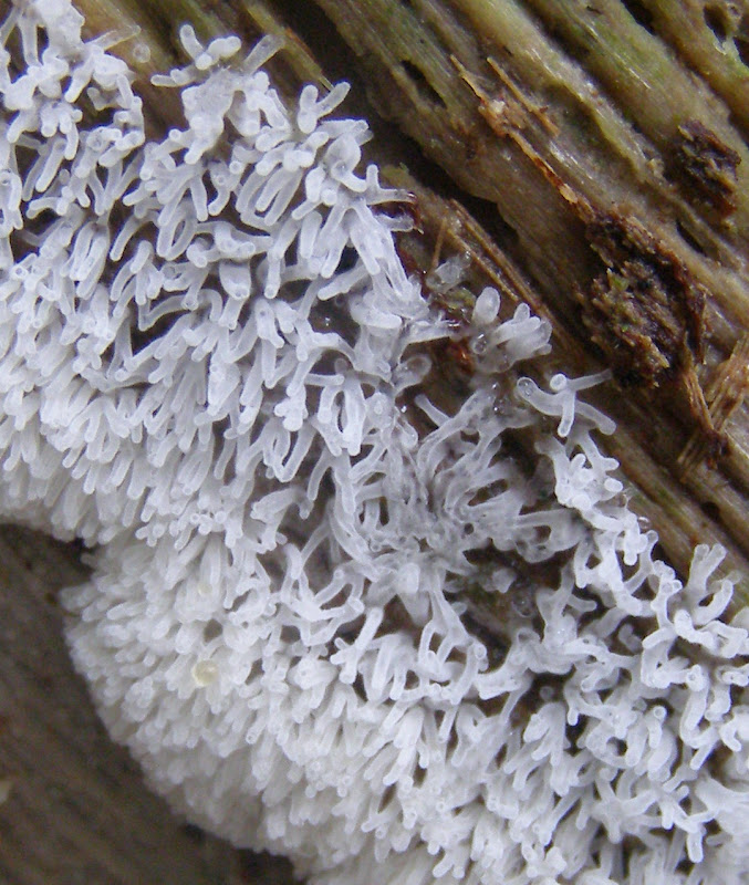
Slime Mould

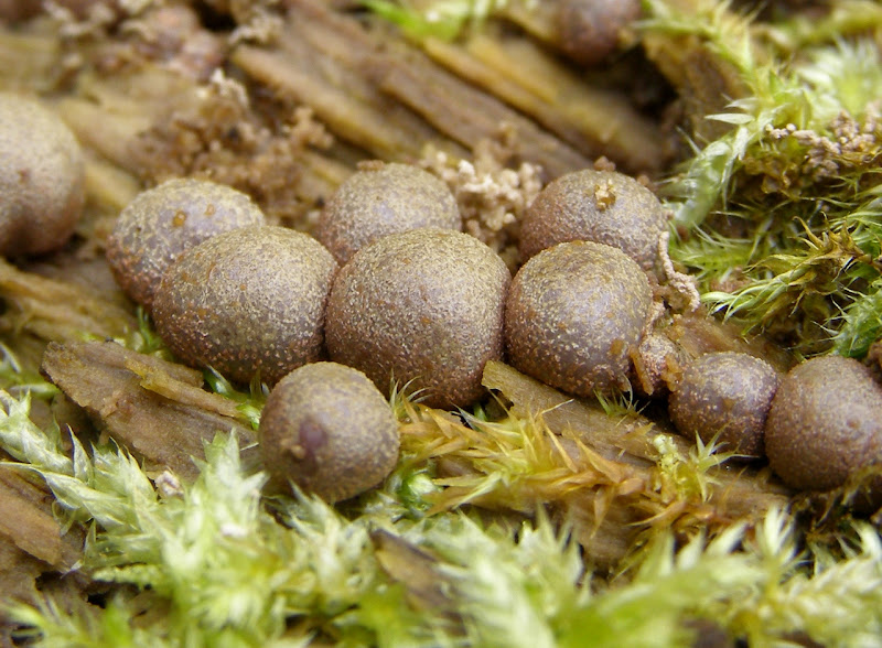
The poroid form of Ceratiomyxa fruticulosa.
See here and here for photos of the filamentous form from last year.

Photos taken in the Wilderness, Whiteknights Park, Reading, UK, on 2013-09-21.
The poroid form of Ceratiomyxa fruticulosa.
See here and here for photos of the filamentous form from last year.
Photos taken in the Wilderness, Whiteknights Park, Reading, UK, on 2013-09-21.

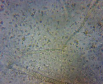
Fruiting bodies of the slime mould Lycogala epidendrum. These fruiting bodies are know as aethalia and are much larger than the smaller sporangia of most other slime moulds.
The older aethalia are brown.
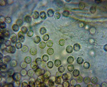
The spores are spherical and are about 6um in diameter (this x600 image is about 86um in width). This is consistent with the 6 - 8um given for Lycogala epidendrum by Stephenson and Stempen (Myxomycetes: A Handbook of Slime Molds, 1994, page 136). You can also see some of the tubular pseudocapillitium in the background.
First 2 photos taken in the Wilderness, Whiteknights Park, Reading, UK, on 2012-05-06.
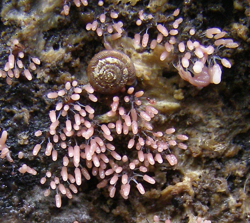
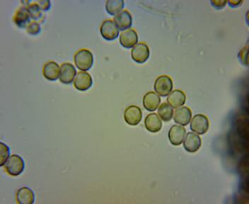
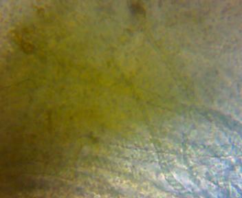
A slime mould with pale pink cylindrical sporangia on stalks, probably Stemonitopsis typhina (=Comatrichia typhoides). The sporangia of this species start out as white but rapidly (within only a day or two) turn pink, then brown and black. For some of the same species that I saw last year, see here.
This is what the spores looked like at x600 under the microscope:
First photo taken in the Wilderness, Whiteknights Park, Reading, UK, on 2012-05-11.
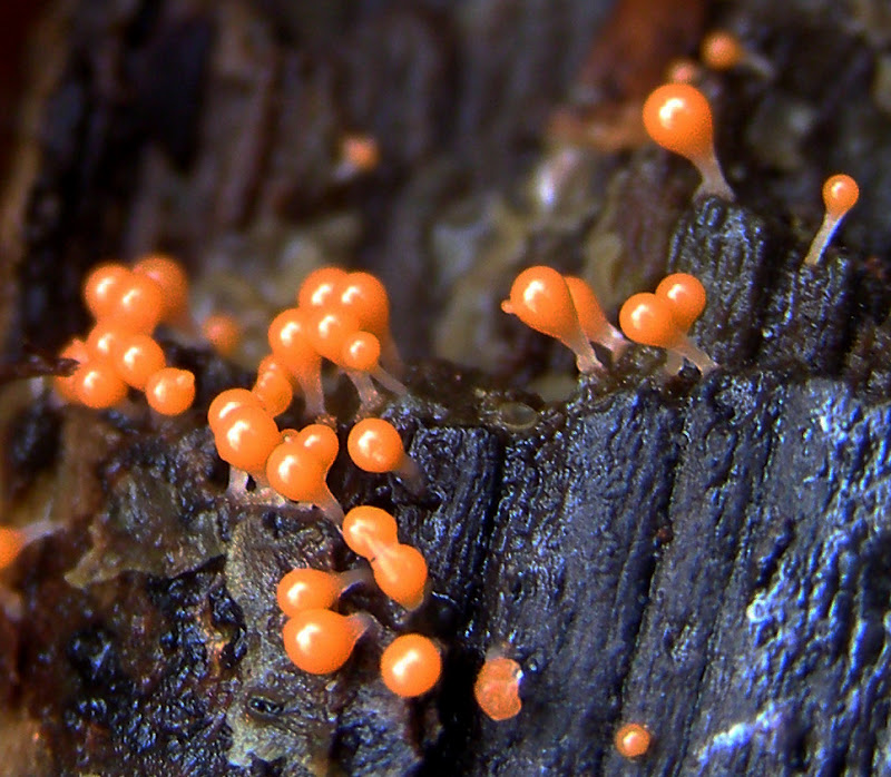
Orange slime mould fruiting bodies growing on decaying wood.
My microscope at x600 shows that the capillitium of these fruiting bodies consists of twisted strands with long tapered ends. This, combined with the orange colour, suggest the species is Trichia decipiens.
First photo taken in the Wilderness, Whiteknights Park, Reading, UK, 2012-11-30.
Slime mould Ceratiomyxa fruticulosa growing on a decaying log.
Photo taken in the Wilderness, Whiteknights Park, Reading, UK, on 2012-10-24.
 Tristram Brelstaff
